Eye Anatomy: The Parts of the Human Eye Explained
A clear guide to eye anatomy — the cornea, iris, lens, retina, and optic nerve. Learn what each part does and how to read and label an eye diagram.
The human eye is often compared to a camera, but that comparison undersells it. In a space smaller than a ping-pong ball, the eye gathers light, focuses it with two lenses, controls its own exposure, and converts the result into electrical signals — all continuously and automatically. Understanding eye anatomy means understanding how a handful of transparent and light-sensitive structures work together to produce vision. This guide walks through every major part of the eye, what each one does, and how to read and label an eye anatomy diagram with confidence.
Quick Answer: What Is the Anatomy of the Eye?
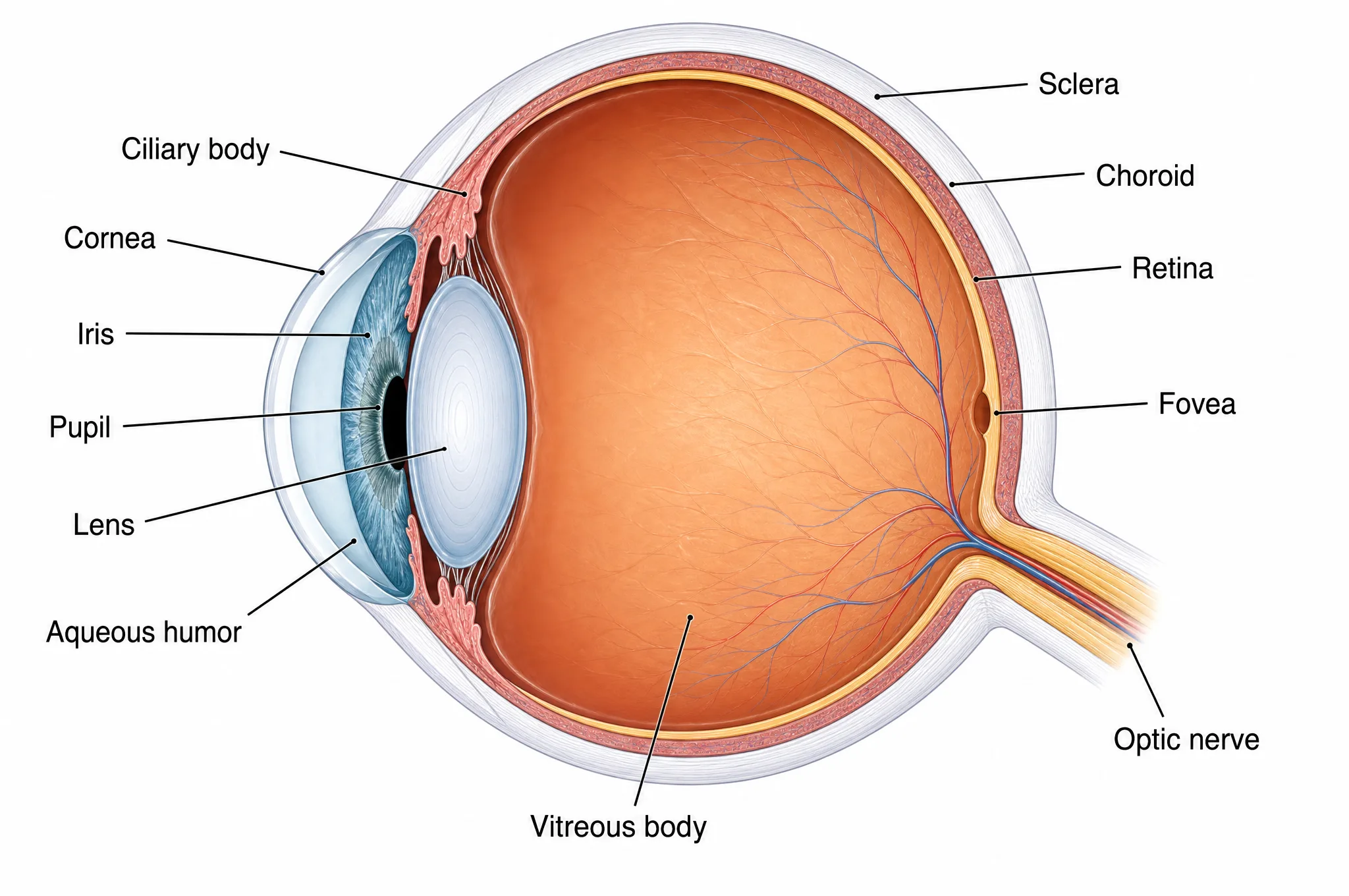
The eye is a roughly spherical organ built from three concentric layers. The outer layer is the tough white sclera, which becomes the clear cornea at the front. The middle layer, called the uvea, contains the choroid, ciliary body, and iris, and supplies blood and controls light. The inner layer is the retina, the light-sensitive tissue that lines the back of the eye.
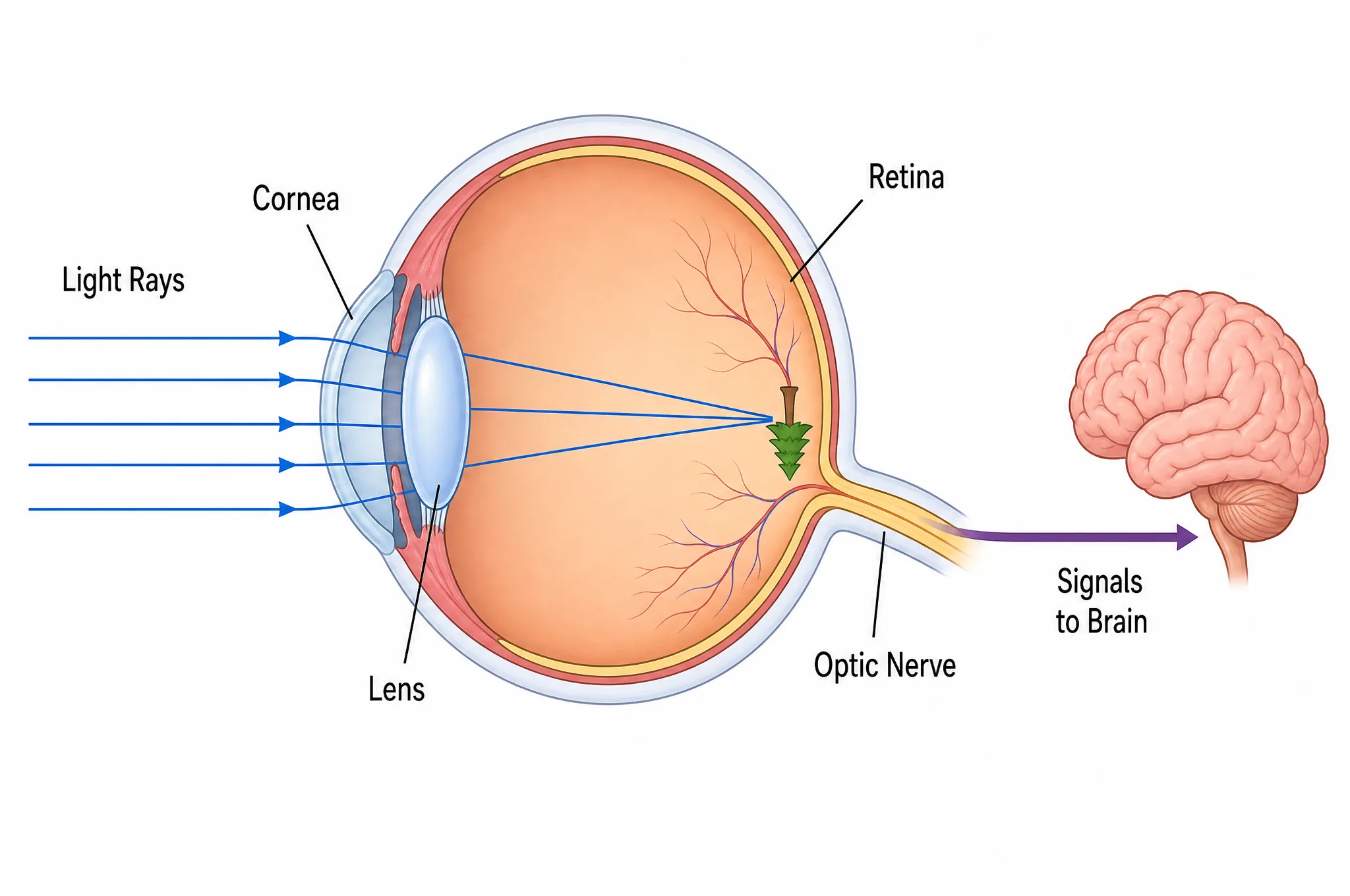
Light enters through the cornea, passes through the pupil and lens, travels across the gel-filled interior, and lands on the retina. There, photoreceptor cells convert light into nerve signals that the optic nerve carries to the brain. Every labeled eye diagram is really just a map of that journey.
The Three Layers of the Eyeball
Almost every structure in the eye belongs to one of three layers, so learning the layers first makes the rest of the anatomy click into place.
- The fibrous layer (outer). This is the eye's protective shell. Most of it is the opaque white sclera — the "white of the eye" — which holds the eyeball's shape and anchors the muscles that move it. At the very front, the fibrous layer becomes transparent and bulges slightly outward as the cornea.
- The vascular layer (middle), or uvea. Rich in blood vessels, this layer nourishes the eye and manages light. It includes the choroid (a dark, blood-rich sheet behind the retina), the ciliary body (which holds and shapes the lens and produces fluid), and the iris (the colored ring that adjusts the pupil).
- The inner layer. This is the retina, the neural tissue where vision actually begins.
A common point of confusion is that the cornea and sclera are the same layer, just transparent in one place and opaque in another. Keeping that in mind prevents a lot of labeling errors.
The Front of the Eye: Cornea, Iris, Pupil, and Lens
The front of the eye is where light is collected and metered before it ever reaches the retina.
Cornea. The clear, dome-shaped front window of the eye. Because light bends sharply when it crosses from air into the curved, denser cornea, the cornea provides roughly two-thirds of the eye's total focusing power. It has no blood vessels — that is part of why it stays perfectly clear — and gets its oxygen directly from the air and from the fluid behind it.
Aqueous humor. Behind the cornea sits a watery fluid called the aqueous humor, which fills the anterior chamber. The ciliary body continuously produces it, and it drains away at a matching rate to keep the pressure inside the eye stable. This fluid also delivers nutrients to the cornea and lens.
Iris and pupil. The iris is the colored part of the eye, a ring of muscle that opens and closes the pupil — the black opening at its center. In bright light the iris constricts the pupil to let in less light; in dim conditions it dilates the pupil to let in more. The pupil only looks black because you are seeing into the dark interior of the eye.
Lens and ciliary body. Just behind the iris is the lens, a transparent, flexible disc suspended by fine fibers from the surrounding ciliary body. While the cornea does the bulk of the focusing, the lens supplies the remaining power and — crucially — can change shape to fine-tune focus for objects at different distances.
The Inside and Back: Retina, Macula, and Optic Nerve
Past the lens, light crosses the large interior of the eye and reaches the structures that actually capture the image.
Vitreous humor. The big cavity between the lens and the retina is filled with the vitreous humor, a clear jelly-like substance that helps the eyeball hold its round shape and keeps the retina pressed smoothly against the back wall.
Retina. Lining the back of the eye, the retina is a thin sheet of neural tissue containing the eye's photoreceptors. These cells convert light into electrical signals. There are two kinds: rods, which number about 120 million, are extremely sensitive to dim light, and handle night and peripheral vision in shades of gray; and cones, about 6 million of them, which work in bright light and detect color.
Macula and fovea. Near the center of the retina is the macula, a small region specialized for detailed central vision. At its very center sits the fovea, a tiny pit packed with the highest density of cones anywhere in the eye. When you read this sentence, you are pointing your fovea at the words — it provides your sharpest, most color-rich vision.
Optic nerve and the blind spot. All the signals from the retina gather and exit the back of the eye through the optic nerve, a bundle of more than a million nerve fibers leading to the brain. The exact spot where the nerve leaves — the optic disc — has no photoreceptors, which creates a natural blind spot. You never notice it in daily life because your brain fills the gap using your other eye and the surrounding scene.
Choroid. Sandwiched between the retina and the sclera, the choroid is a dark, blood-rich layer that nourishes the outer retina and absorbs stray light to keep the image crisp.
How the Eye Focuses Light
Vision depends on bending incoming light so that it converges precisely on the retina. The eye does this in two stages.
First, the cornea performs most of the bending. Because the difference in optical density between air and corneal tissue is large, light refracts strongly the moment it enters — supplying about two-thirds of the eye's focusing power as a fixed, always-on lens.
Second, the lens fine-tunes the focus through a process called accommodation. To see something close up, the ciliary muscle contracts, the suspending fibers slacken, and the elastic lens becomes rounder and more powerful. To see something far away, the muscle relaxes, the fibers pull the lens flatter, and its focusing power drops. This constant reshaping is what lets you glance from a far wall to a nearby page without anything appearing blurry.
When this two-stage system is slightly off — for example, if the eyeball is a little too long — light focuses in front of the retina instead of on it, producing nearsightedness. Many common vision problems are, at heart, focusing-geometry problems.
Why Eye Anatomy Matters
Knowing the parts of the eye is not just trivia. It is the vocabulary that doctors, students, and designers use to talk about vision.
- In medicine, conditions are named for the structures they affect: cataracts cloud the lens, glaucoma involves pressure from aqueous humor, macular degeneration damages the fovea-bearing macula, and retinal detachment lifts the retina off the back wall. You cannot follow any of these without the anatomy.
- In learning, an accurate mental map of the eye makes the physics of light and the biology of perception far easier to understand.
- In design and communication, a well-labeled eye diagram is one of the most-used illustrations in biology, optometry, and science education.
How to Read and Label an Eye Diagram
A standard eye diagram is a side-on cross-section, as if the eyeball were sliced in half from front to back. Reading it is straightforward once you know the route light takes.
- Start at the front and follow the light. Label the cornea first, then the aqueous humor behind it, then the iris and pupil, then the lens and ciliary body.
- Cross the middle. The large central space is the vitreous humor.
- Work outward at the back. From the inside out, the three back-wall layers are retina, then choroid, then sclera. Mark the macula and fovea on the retina near the central axis.
- Finish with the exit. Label the optic nerve where it leaves the back of the eye.
Two habits prevent most mistakes: always label layers from a single consistent direction (inside-out or outside-in), and keep your leader lines from crossing, which keeps a busy diagram readable.
Common Mistakes and Confusions
- Confusing the cornea and the lens. Both are transparent and both focus light, but the cornea is the fixed front window that does most of the work, while the lens sits behind the iris and adjusts focus.
- Treating the cornea and sclera as different layers. They are the same fibrous layer — clear at the front, white everywhere else.
- Mixing up the two fluids. The watery aqueous humor is in the small front chamber; the jelly-like vitreous humor fills the large rear cavity.
- Swapping rods and cones. Rods are the many dim-light, gray-scale cells; cones are the fewer bright-light, color cells concentrated at the fovea.
- Forgetting the choroid. Many beginners label retina and sclera but skip the dark, blood-rich choroid layer between them.
Frequently Asked Questions
- What are the main parts of the eye?
- The main parts are the cornea, iris, pupil, lens, and ciliary body at the front; the sclera and choroid forming the outer and middle walls; and the retina, macula, fovea, and optic nerve at the back. The aqueous humor fills the front chamber and the vitreous humor fills the large rear cavity.
- What is the difference between rods and cones?
- Rods and cones are the retina's two photoreceptor types. Rods are far more numerous (about 120 million), are very sensitive to dim light, and handle night and peripheral vision in shades of gray. Cones (about 6 million) work in bright light, detect color, and are packed most densely in the fovea for sharp central vision.
- How does the eye focus light?
- The cornea does most of the focusing — roughly two-thirds of the eye's refractive power — because light bends sharply when it passes from air into the curved cornea. The lens supplies the rest and fine-tunes focus by changing shape, a process called accommodation, so you can switch between distant and near objects.
- What is the macula and the fovea?
- The macula is a small, specialized region near the center of the retina responsible for detailed central vision. At its center lies the fovea, a tiny pit with the highest density of cones in the eye, which gives you your sharpest, most color-rich vision.
- Why does the eye have a blind spot?
- The optic disc is the point where the optic nerve and blood vessels leave the eye. It has no photoreceptors, so no image forms there — creating a natural blind spot. You normally never notice it because your brain fills in the gap using the other eye and surrounding visual information.